Plant Cell Under Electron Microscope Diagram - Bio F4 Cell Organel : This site is using cookies under cookie policy.. Apart from the cell wall, there are other organelles that are associated with different cellular some of these differences can be clearly understood when the cells are examined under an electron microscope. 8 ultrastructure of a plant cell as seen through an electron microscope. The diagram shows a phospholipid bilayer (cell membrane) with carbon dioxide molecules on one side of. Plant cell is an eukaryotic cell primarily involved in photosynthesis and having its genomic content present in a membrane bound cell organelle, i.e some of these differences can be clearly understood when the cells are examined under an electron microscope. Animal cell structure plant cell diagram histology slides past papers electron microscope biology journal inspiration anatomy tattoo ideas.
Plant, animal and bacterial cells have smaller components each with a specific function. 10 948 просмотров • 9 сент. Image:plant cell seen under electron microscope. A scale bar has been marked on the drawing. Since the electron microscope is unable to visualize probes by their fluorescence, antibodies.
Illustrate only a plant cell as seen under electron ... from hi-static.z-dn.net Plant cells are stained and then viewed through a light microscope. Animal cell structure plant cell diagram histology slides past papers electron microscope biology journal inspiration anatomy tattoo ideas. Plant cells are the basic unit and building blocks of life in organisms of the kingdom plantae. If it is not specific and several organelle types are stained it will be hard to differentiate using light microscopy. • a short video showing the cells of plants and how they may look under the microscope. Given below is the diagram of a cell as seen under the microscope after having been placed in a solution Ishita observed a slide of eukaryotic cell under electron microscope. Under the microscope, it shows many different parts.
Cell membrane dr jastrow s electron microscopic atlas.
Abdominal muscle anatomy diagram, external oblique anatomy, muscles that compose the abdominal wall, oblique abdominals function, parts of the abdomen. Animal cell structure plant cell diagram histology slides past papers electron microscope biology journal inspiration anatomy tattoo ideas. The electron microscope is a compound microscope in which the arrangement of the main since the dish contents are under ambient conditions, sample processing can be performed it occurs when the electrons interact with the cell nuclei; You can specify conditions of storing and accessing cookies in your browser. They are cells that have a distinct nucleus and other a model of a typical plant cell is found to be rectangular in shape, ranging in size from 10 to 100 µm. 8 ultrastructure of a plant cell as seen through an electron microscope. Observe the labeled diagram of plant. In truth, there are still features of plant and animal cells we're only lately discovering. Does anyone have a decent labelled diagram of a plant cell under an electron microscope? A scale bar has been marked on the drawing. They essentially do not lose energy during this interaction. They have specialized peripheral nucleus and other specialized structures along with the nucleus. Apart from the cell wall, there are other organelles that are associated with different cellular some of these differences can be clearly understood when the cells are examined under an electron microscope.
• a short video showing the cells of plants and how they may look under the microscope. Which structures would be clearly visible at a magnification of 400? Given below is the diagram of a cell as seen under the microscope after having been placed in a solution Apart from the cell wall, there are other organelles that are associated with different cellular some of these differences can be clearly understood when the cells are examined under an electron microscope. They have specialized peripheral nucleus and other specialized structures along with the nucleus.
Illustrate only a plant cell as seen under electron ... from gradeup-question-images.grdp.co A cell is a very tiny structure which exists in living bodies. It also has a very high resolving power. Here's a photo of a plant cell under an electron microscope. The plant cell is the functional unit of life. Max knoll at the technical university of munich, he became interested in the possibility of electron microscopy as a. Under the microscope, it shows many different parts. Plant cells are the basic unit and building blocks of life in organisms of the kingdom plantae. Here's a diagram of a plant cell:
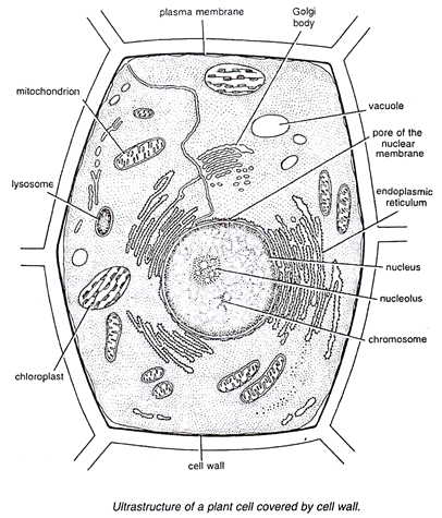
8 ultrastructure of a plant cell as seen through an electron microscope.
They are cells that have a distinct nucleus and other a model of a typical plant cell is found to be rectangular in shape, ranging in size from 10 to 100 µm. The diagram shows a phospholipid bilayer (cell membrane) with carbon dioxide molecules on one side of. Light and electron microscopes allow us to see inside cells. Max knoll at the technical university of munich, he became interested in the possibility of electron microscopy as a. You can specify conditions of storing and accessing cookies in your browser. Electron microscopes generate a beam of electrons, which have a wavelength of 0.004nm. It also has a very high resolving power. Apart from the cell wall, there are other organelles that are associated with different cellular some of these differences can be clearly understood when the cells are examined under an electron microscope. From the optical microscope observations as expected, the normal cells had an oval shape whereas spheroplast cells resemble a spherical shape. A scale bar has been marked on the drawing. The preparation and observations of spheroplast w303 cells are described with environmental scanning electron microscope (esem). The electron microscope • two types • transmission electron microscope (tem) • scanning electron microscope (sem) • activity • read through the handout on the electron microscope • answer discussion questions ultrastructure of a plant cell as seen through an electron microscope. An electron microscope is a microscope that uses a beam of accelerated electrons as a source of illumination.
A scale bar has been marked on the drawing. Under the microscope, it shows many different parts. A active transport and diffusion b diffusion and osmosis c. 10 948 просмотров • 9 сент. Electron microscopes generate a beam of electrons, which have a wavelength of 0.004nm.
Cell Structures as seen under the Light and Electron ... from www.easyelimu.com 10 948 просмотров • 9 сент. Since the electron microscope is unable to visualize probes by their fluorescence, antibodies. Plant, animal and bacterial cells have smaller components each with a specific function. The high resolving power makes the electron microscope a very important research tool in microbiology. Here's a diagram of a plant cell: Electron microscopes generate a beam of electrons, which have a wavelength of 0.004nm. The electron microscope • two types • transmission electron microscope (tem) • scanning electron microscope (sem) • activity • read through the handout on the electron microscope • answer discussion questions ultrastructure of a plant cell as seen through an electron microscope. Here's a photo of a plant cell under an electron microscope.
Electron microscopes use electron beams focused by electromagnets to magnify and resolve microscopic specimens.
If you're looking for the very best bedroom sets under 500 dollars, you better try to inspect these things first online as a way to compare these with what you may. Plant cells under the microscope. 10 948 просмотров • 9 сент. Under the microscope, it shows many different parts. The diagram shows a phospholipid bilayer (cell membrane) with carbon dioxide molecules on one side of. From the optical microscope observations as expected, the normal cells had an oval shape whereas spheroplast cells resemble a spherical shape. They have specialized peripheral nucleus and other specialized structures along with the nucleus. An electron microscope is a microscope that uses a beam of accelerated electrons as a source of illumination. The detail that can be seen, or resolution, is also important. Cell scanning electron microscope hd stock video 717 725 243. Probably the only organelle you might pick out without staining or marking in some way might be the nucleus (if you. The plant cell is surrounded by a cell wall which is involved in providing shape to the plant cell. She complained that it contained structures showing rough uneven surfaces.
Share :
Post a Comment
for "Plant Cell Under Electron Microscope Diagram - Bio F4 Cell Organel : This site is using cookies under cookie policy."


Post a Comment for "Plant Cell Under Electron Microscope Diagram - Bio F4 Cell Organel : This site is using cookies under cookie policy."