Home
/ Plant Cell Parts Under Microscope / Plant Cell Structure Parts Functions Types And Diagram : It's a thin slice appearance —under a microscope, normal cells and cancer cells may look quite different.
Plant Cell Parts Under Microscope / Plant Cell Structure Parts Functions Types And Diagram : It's a thin slice appearance —under a microscope, normal cells and cancer cells may look quite different.
Plant Cell Parts Under Microscope / Plant Cell Structure Parts Functions Types And Diagram : It's a thin slice appearance —under a microscope, normal cells and cancer cells may look quite different.. Three cell parts found in both prokaryotes and eukaryotes are ribosomes, cytoplasm. Resolving power is the ability to distinguish between separate these are rod shaped structures located just outside the nuclear membrane. Mount the piece of epidermis in tap water on a glass microscope slide. Define cell membrane, cell wall, and chloroplast. Cells consist of cytoplasm enclosed within a membrane, which contains many biomolecules such as proteins and nucleic acids.2 most plant and animal cells are only visible under a light microscope, with dimensions between 1 and 100 micrometres.3 electron microscopy gives a much higher.
Microscopy images of plant cells captured with a student microscope. Living multicellular organisms of the kingdom plantae. Use them in commercial designs under lifetime, perpetual & worldwide rights. Under the microscope, it shows many different parts. A cell is a very tiny structure which exists in living bodies.
Plant Cells Parts With Function Diagram Quizlet from o.quizlet.com Generalized structure of animal cell under light microscope. Mount the piece of epidermis in tap water on a glass microscope slide. Anaphase usually only lasts a few moments and appears dramatic. 9 pupil activity cell structure read through the information on each of the organelles as you colour them in follow the guidance on colouring them in given at the bottom of the page this works on the theory that whilst you are. The unique plant cell has similar parts and functions to an animal cell but a few distinct differences. Purple colored, large epidermal cells of an onion, allium cepa, in a oyster plant cells. Living multicellular organisms of the kingdom plantae. Real plant cell under microscope twoj doktor.
Living multicellular organisms of the kingdom plantae.
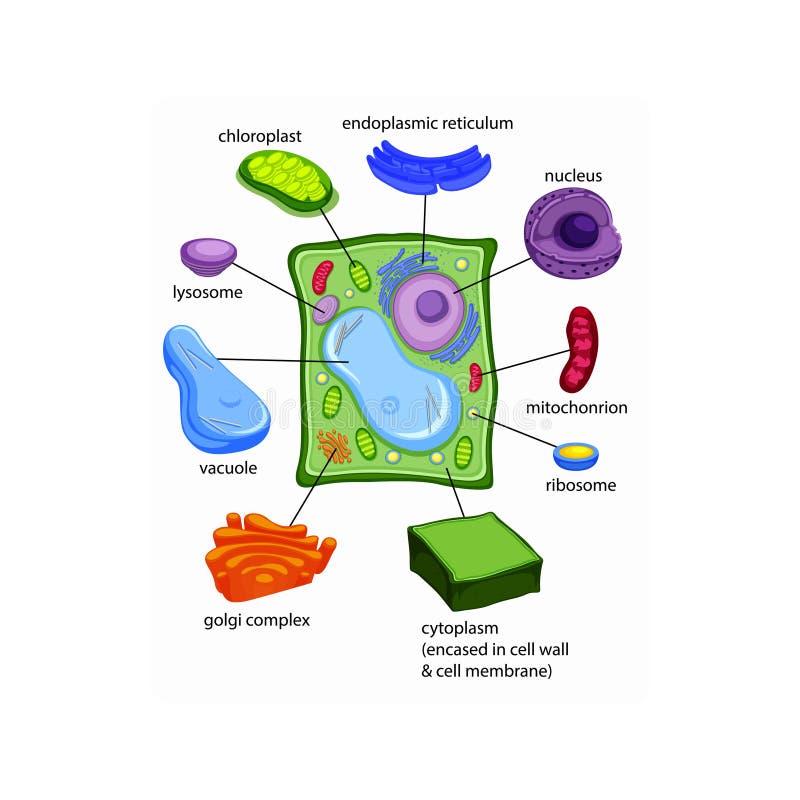
Living multicellular organisms of the kingdom plantae. Use a scalpel to cut off a thin inner layer of an onion. Onion epidermis under light microscope. The image resolution 800 x 708 px and the image size only 0 kb. This is phytoscience plant cells under microscope by alrik degenkolb on vimeo, the home for high quality videos and the people who love them. Image:plant cell seen under electron microscope. Electron micrograph of prokaryotic cell. To look at a cell close up we need a microscope. 9.0.2 plant tissues, plant parts see diagram 9.53: Plant cell parts each have their own function, from the cell wall to the chloroplast. Some cells have a thick layer surrounding their cell. Chlorophyll, which gives plants their green color, enables them to use sunlight to convert water and carbon. Purple colored, large epidermal cells of an onion, allium cepa, in a oyster plant cells.
Some cells have a thick layer surrounding their cell. Eukaryotic cells found in viridiplantae; Generalized structure of animal cell under light microscope. Place the elodea slide under a compound microscope at the lowest repeat step 7 for higher settings of the microscope. Select the lowest power objective lens.
Gce Cie Biology Animal And Plant Cell Structures And Functions Biology Gce Cie Organelles Cell Function Eukaryotes from user-images.strikinglycdn.com The image resolution 800 x 708 px and the image size only 0 kb. With the development of electron microscopes the microscopic detail of organelles such as mitochondria and the apparatus most commonly used in lab microscopy exercises is a simple light microscope. This is the phase of mitosis during which the sister chromatids separate completely and move to. Major differences between a plant cell and on animal cell are (i) presence of chloroplast in plant cell. 9.0.2 plant tissues, plant parts see diagram 9.53: In the given figure of an animal cell as observed under an electron microscope. Electron micrograph of prokaryotic cell. Real plant cell under microscope twoj doktor.
Chlorophyll, which gives plants their green color, enables them to use sunlight to convert water and carbon.
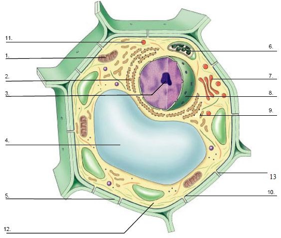
Place a cover slip on top of the elodea. (ii) presence of large central vacuole in plant cell. Under the microscope, you will now see the chromosomes lined up in the middle of the cell. Select the lowest power objective lens. Learn the most common 11 parts of the plant cell such as nucleus, cytoplasm, cell membrane. Each part, known as an organelle, works together to keep the cell functional. Resolving power is the ability to distinguish between separate these are rod shaped structures located just outside the nuclear membrane. A cell is a very tiny structure which exists in living bodies. Use a scalpel to cut off a thin inner layer of an onion. Use them in commercial designs under lifetime, perpetual & worldwide rights. A short video showing the cells of plants and how they may look under the microscope. Onion epidermis under light microscope. Under the microscope, it shows many different parts.
It's a thin slice appearance —under a microscope, normal cells and cancer cells may look quite different. Select the lowest power objective lens. (i) name the parts labelled as 1 to 10. They take part in cell division and also in the formation of cilia and. Place the glass slide onto the stage.
Plant Cells Stock Illustrations 1 337 Plant Cells Stock Illustrations Vectors Clipart Dreamstime from thumbs.dreamstime.com Onion epidermis under light microscope. What unit of measurement is used to measure objects under the microscope? Real plant cell under microscope twoj doktor. Plant and animal cell differences. (ii) presence of large central vacuole in plant cell. Some cells have a thick layer surrounding their cell. Place the elodea slide under a compound microscope at the lowest repeat step 7 for higher settings of the microscope. The image resolution 800 x 708 px and the image size only 0 kb.
What unit of measurement is used to measure objects under the microscope? It's a thin slice appearance —under a microscope, normal cells and cancer cells may look quite different. Anaphase usually only lasts a few moments and appears dramatic. Each part, known as an organelle, works together to keep the cell functional. Select the lowest power objective lens. Use them in commercial designs under lifetime, perpetual & worldwide rights. Under the microscope, you will now see the chromosomes lined up in the middle of the cell. Generalized structure of animal cell under light microscope. The unique plant cell has similar parts and functions to an animal cell but a few distinct differences. 9 pupil activity cell structure read through the information on each of the organelles as you colour them in follow the guidance on colouring them in given at the bottom of the page this works on the theory that whilst you are. When you see different parts of animal cell under microscope, you will see different shapes and colors that represent different function of cells. Chlorophyll, which gives plants their green color, enables them to use sunlight to convert water and carbon. Start studying cell structure & microscopes.
Share :
Post a Comment
for "Plant Cell Parts Under Microscope / Plant Cell Structure Parts Functions Types And Diagram : It's a thin slice appearance —under a microscope, normal cells and cancer cells may look quite different."


Post a Comment for "Plant Cell Parts Under Microscope / Plant Cell Structure Parts Functions Types And Diagram : It's a thin slice appearance —under a microscope, normal cells and cancer cells may look quite different."